A condensed guide for PT students and new grads. Seventeen shoulder conditions, each in four steps: recognize it, confirm it, differentiate it, and treat it.
← All Clinical RoadmapsTap any roadmap and it opens right here. Every route makes the same four stops, so you always know where you are.
Thinking Clinically · A condensed guide for PT students and new grads
Rotator cuff-related shoulder pain (RCRSP) is one of the most common shoulder diagnoses you will see. It is a clinical syndrome, not a single structural diagnosis, so the useful question is not “which tendon is torn?” but “does this presentation fit a load-sensitive shoulder?” This roadmap walks through how to recognize it, confirm it, rule out its mimics, and rebuild the shoulder’s capacity to tolerate load.
- Pain in the lateral or anterior shoulder, sometimes into the upper arm, usually with gradual onset tied to a change in activity or load
- Symptoms driven by load: overhead reaching, lifting, pushing, pulling, and sustained use
- Trouble with dressing, reaching behind the back, grooming, and sleeping on the involved side
- Painful active motion with relatively normal passive motion, and a possible painful arc that doesn’t confirm the diagnosis on its own
- Reduced strength that may reflect pain inhibition rather than a structural tear, and a clean neuro screen
- No single special test carries enough weight to stand alone. You’re building a cluster
- History: gradual onset after a change in shoulder use, aching or catching with specific movements, and symptoms that fluctuate with activity volume. Ask what is hard to do, because functional triggers tell you as much as a special test.
- AROM that is full but painful, especially through a painful arc, with PROM relatively preserved. That gap is one of your most useful signals.
- Resisted external rotation, Jobe/full can, and resisted abduction reproduce familiar pain, sometimes with mild weakness.
- Scapular movement may look altered, but don’t assume it causes the pain. Ask whether changing the scapular strategy changes the symptoms.
- Diagnostic value comes from concordance: history, painful arc, resisted loading response, and no competing findings all pointing the same direction.
| Adhesive capsulitis | Both AROM and PROM restricted, especially external rotation, with stiffness even without much loading |
|---|---|
| Glenohumeral OA | Older adult with global motion loss, crepitus, and symptoms not specifically tied to cuff loading |
| AC joint pain | Pain squarely over the AC joint, reproduced by cross-body adduction |
| Cervical radiculopathy | Pain past the elbow, numbness or tingling, dermatomal, myotomal, or reflex change, or neck movement reproducing arm symptoms |
| Non-musculoskeletal causes | Systemic symptoms, unexplained weight loss, chest symptoms, or significant trauma with inability to actively elevate the arm |
Pain that doesn’t match the mechanical pattern, new neurological findings, systemic symptoms, or significant trauma with inability to lift the arm all warrant a closer look before you stay in the RCRSP lane.
- Start with education. Patients often assume pain equals damage. A useful frame: the shoulder has become less tolerant of the load it’s being asked to handle, not more fragile, and the job is to rebuild that tolerance.
- Match activity modification to irritability instead of blanket rest. High irritability means reducing volume and range, not full avoidance.
- Use manual therapy as a bridge, not the destination, when it creates a short-term change that lets the patient move and load better.
- Phase 1: establish load tolerance with ER/IR isometrics, supported elevation, and scapular control, dosed to irritability.
- Phase 2: build strength with resisted ER/IR, scaption, rows, and serratus work, changing one or two variables at a time.
- Phase 3: restore capacity with overhead pressing, carries, loaded reaching, and work- or sport-specific demands. Progress by tolerable symptoms and improving function, not a pain-free session.
RCRSP rewards clinicians who think in patterns instead of chasing a single positive test. When the history, painful elevation, resisted loading response, and functional limits tell a consistent story, and you’ve reasonably screened out the differentials, you have a strong working diagnosis: progressively rebuild what the shoulder can tolerate.
Watch exercise demonstrations and clinical reasoning videos on the Thinking Clinically YouTube channel: youtube.com/@Thinking.Clinically
- Desmeules F, Roy JS, Lafrance S, et al. Rotator Cuff Tendinopathy Diagnosis, Nonsurgical Medical Care, and Rehabilitation: A Clinical Practice Guideline. Journal of Orthopaedic & Sports Physical Therapy. 2025;55(4):235–274.
- Lafrance S, Charron M, Dubé MO, et al. The Efficacy of Exercise Therapy for Rotator Cuff-Related Shoulder Pain According to the FITT Principle: A Systematic Review With Meta-analyses. JOSPT. 2024;54(8):499–512.
- Tena-Portilla A, Martín-San Agustín R, Gacto-Sánchez M, Escriche-Escuder A. Effectiveness of exercise in rotator cuff-related shoulder pain: a systematic review with meta-analysis. Physiotherapy Theory and Practice. 2026.
- Zhang B, Raguzzi IA, Dupuis F, et al. Addressing Shoulder Weakness in Individuals With Rotator Cuff-Related Shoulder Pain: A Systematic Review With Meta-analysis. JOSPT. 2026;56(2):67–84.
- Hughes PC, Taylor NF, Green RA. Most clinical tests cannot accurately diagnose rotator cuff pathology: a systematic review. Australian Journal of Physiotherapy. 2008;54(3):159–170.
- Longo UG, Berton A, Ahrens PM, et al. Clinical tests for the diagnosis of rotator cuff disease. Sports Medicine and Arthroscopy Review. 2011;19(3):266–278.
- Lafrance S, Charron M, Roy JS, et al. Diagnosing, Managing, and Supporting Return to Work of Adults With Rotator Cuff Disorders: A Clinical Practice Guideline. J Orthop Sports Phys Ther. 2022;52(10):647–664.
Thinking Clinically · A condensed guide for PT students and new grads
A rotator cuff tear is a partial- or full-thickness disruption of one or more cuff tendons, most often the supraspinatus. A tear on imaging does not automatically equal the pain generator, so the clinical question is whether the tear is contributing to pain, weakness, or loss of function. This roadmap covers the symptomatic, nonoperative presentation. Postoperative care is in the surgical roadmaps.
- Middle-aged or older adult (traumatic tears can occur at any age) with lateral or anterolateral shoulder or upper-arm pain
- Pain with reaching, lifting, pushing, pulling, and overhead activity, and trouble sleeping on the involved side
- Weakness that is greater than expected from pain alone
- Reduced active elevation with relatively preserved passive ROM, and difficulty holding the arm against gravity or controlling lowering
- Possible lag signs; chronic tears may show atrophy or compensatory movement
- A fall, sudden pull, or heavy catch followed by immediate weakness or inability to elevate the arm deserves medical evaluation and imaging
- The separator question: is the patient unable to move because it hurts, or unable to move because they cannot generate or control force?
- AROM much worse than PROM means think force production before you think mobility.
- Build a cluster: external rotation lag sign, substantial ER weakness not explained by pain, Jobe/full can weakness, and drop arm or inability to control elevation. One positive test is a clue; several concordant findings make a pattern.
- Screen the subscapularis with belly-press, lift-off, and bear-hug. No single subscapularis test is sensitive enough to exclude a tear.
- Clinical tests cannot reliably determine tear size. Consider imaging when the result will change management, especially after a substantial traumatic event.
| Rotator cuff-related shoulder pain | Pain is the main limiter, strength is relatively preserved, AROM is near full, and there is no lag |
|---|---|
| Adhesive capsulitis | Both AROM and PROM restricted, especially external rotation, with stiffness more prominent than weakness |
| Glenohumeral OA | AROM and PROM both restricted, with joint-line pain, crepitus, or mechanical symptoms |
| Cervical radiculopathy | Neck pain, symptoms beyond the shoulder, numbness or tingling, myotomal or reflex change |
| Proximal humerus fracture | Significant trauma, severe immediate pain, unable to use the arm, focal bony tenderness, deformity or bruising |
A traumatic event followed by major weakness, inability to actively elevate, or pseudoparalysis should not go into routine rehab without considering medical evaluation and imaging. Progressive weakness and new neurological findings are also reasons to reassess.
- Educate first. A tear on imaging doesn’t automatically explain pain, and the goal is to improve how the shoulder works, not to make the image look normal. For many symptomatic small-to-medium full-thickness tears, the 2025 AAOS guideline gives strong support for either physical therapy or surgery in appropriate patients.
- Modify activity instead of prescribing complete rest. Reduce heavy overhead lifting early when irritability is high, then reintroduce provocative loads gradually. Load is not automatically damage.
- Use manual therapy as an adjunct to active rehabilitation, not a substitute for progressive loading.
- Phase 1: symptom control and movement. Supported or assisted movement, plus ER, IR, abduction, and flexion isometrics (3 to 5 reps of 20 to 30 second holds), dosed to irritability.
- Phase 2: progress to isotonic loading with side-lying and band ER, band IR, supported scaption, rows, and serratus work. Don’t ignore the deltoid, which can matter a lot for elevation when the cuff is compromised.
- Phase 3: functional loading from waist to shoulder to overhead lifting, carries, and work- or sport-specific tasks. Progress because capacity has improved, not because weeks have passed.
- Reassess the plan if weakness is worsening, the patient loses the ability to elevate, pain worsens despite load modification, or progress stalls.
A rotator cuff tear is a structural diagnosis, but rehabilitation is a functional problem. Determine what the patient can and cannot do, how much force they can produce, whether pain or structural weakness is limiting performance, and whether conservative care is still appropriate or medical evaluation is warranted.
Watch exercise demonstrations and clinical reasoning videos on the Thinking Clinically YouTube channel: youtube.com/@Thinking.Clinically
- American Academy of Orthopaedic Surgeons. Management of Rotator Cuff Injuries Evidence-Based Clinical Practice Guideline. August 18, 2025.
- Desmeules F, Roy JS, Lafrance S, et al. Rotator Cuff Tendinopathy Diagnosis, Nonsurgical Medical Care, and Rehabilitation: A Clinical Practice Guideline. J Orthop Sports Phys Ther. 2025;55(4):235-274.
- Hermans J, Luime JJ, Meuffels DE, Reijman M, Simel DL, Bierma-Zeinstra SMA. Does this patient with shoulder pain have rotator cuff disease? The Rational Clinical Examination systematic review. JAMA. 2013;310(8):837-847.
- Hughes PC, Taylor NF, Green RA. Most clinical tests cannot accurately diagnose rotator cuff pathology: a systematic review. Aust J Physiother. 2008;54(3):159-170.
- Lädermann A, Meynard T, Denard PJ, Ibrahim M, Saffarini M, Collin P. Reliable diagnosis of posterosuperior rotator cuff tears requires a combination of clinical tests. Knee Surg Sports Traumatol Arthrosc. 2021;29(7):2118-2133.
- Khatri C, Ahmed I, Parsons H, et al. The Natural History of Full-Thickness Rotator Cuff Tears in Randomized Controlled Trials: A Systematic Review and Meta-analysis. Am J Sports Med. 2019;47(7):1734-1743.
- Piper CC, Hughes AJ, Ma Y, Wang H, Neviaser AS. Operative versus nonoperative treatment for the management of full-thickness rotator cuff tears: a systematic review and meta-analysis. J Shoulder Elbow Surg. 2018;27(3):572-576.
- Shepet KH, Liechti DJ, Kuhn JE. Nonoperative treatment of chronic, massive irreparable rotator cuff tears: a systematic review with synthesis of a standardized rehabilitation protocol. J Shoulder Elbow Surg. 2021;30(6):1431-1444.
Thinking Clinically · A condensed guide for PT students and new grads
Adhesive capsulitis, commonly called frozen shoulder, is not just a stiff shoulder. The hallmark is a global, disproportionate loss of passive motion, especially external rotation, along with pain. It is a clinical diagnosis, and the most practical treatment idea comes from the 2013 Orthopaedic Section guideline: classify tissue irritability first, then match the intensity of stretching, mobilization, and exercise to it.
- Most commonly a middle-aged adult with gradual onset of shoulder pain and stiffness; no single traumatic event is required
- Risk factors include age 40 to 65, female sex, and a prior episode in the other shoulder. Diabetes is associated with a poorer course
- Both AROM and PROM are restricted, with external rotation often disproportionately limited, followed by abduction, flexion, and internal rotation
- Pain that can dominate early, with night pain and difficulty sleeping on the involved side
- Increasing trouble reaching overhead, behind the back, and away from the body
- Neurological findings are typically absent
- The 2013 guideline description: gradual, progressive pain (often night pain and pain at end range) with painful, restricted active and passive ROM in elevation and rotation, present for at least 1 month and plateaued or worsening.
- Passive external rotation loss with the arm at the side and at varying degrees of abduction is a particularly useful finding. In adhesive capsulitis, rotation typically becomes more restricted as the arm moves into abduction.
- There is no validated special-test cluster. Build a pattern: marked passive ER loss, passive restriction in multiple planes, AROM and PROM restricted to a similar degree, and painful end range.
- Reconsider the diagnosis if PROM is essentially normal, radiographs show glenohumeral arthritis, rotation increases rather than decreases from 45° to 90° of abduction, subscapularis palpation reproduces the pain, or neural tension testing reproduces the symptoms.
- Classify irritability before you dose anything: pain over 7, 4 to 6, or under 4 out of 10; consistent, intermittent, or no night pain; pain before end range, at end range, or only with overpressure.
| Rotator cuff-related shoulder pain | PROM is relatively preserved, pain is greater than stiffness, and resisted cuff testing reproduces symptoms |
|---|---|
| Rotator cuff tear | Substantial weakness, AROM much worse than PROM, positive lag signs, or traumatic onset |
| Glenohumeral OA | Joint-line pain, crepitus, mechanically aggravated symptoms, and degenerative change on radiographs |
| Cervical radiculopathy | Neck pain, distal radiation, numbness or tingling, myotomal or reflex change |
| Non-musculoskeletal causes | Systemic or constitutional symptoms, severe unremitting pain, or pain that doesn’t behave mechanically |
Screen the cervicothoracic spine, and consider medical referral for features that suggest neoplasm, fracture, or vascular injury. The guideline also notes that postoperative stiffness is not adhesive capsulitis.
- Educate on the natural course, promote activity modification toward functional pain-free ROM, and match stretching intensity to current irritability. Avoid telling patients the capsule must be “broken loose.”
- High irritability: activity modification, heat or electrical stimulation for pain, grade I to II mobilization in pain-free ranges, and pain-free PROM and AAROM.
- Moderate irritability: moderate-intensity mobilization and gentle-to-moderate stretching into tissue resistance, without post-treatment soreness, plus integrating new motion with scapulohumeral movement and reaching.
- Low irritability: end-range grade III/IV mobilization, progressive sustained stretching, and higher-level functional and recreational movement.
- Reclassify at every visit. Irritability drives dosing; the stage of the condition does not.
- Track outcomes with a validated measure such as the SPADI, DASH, or ASES.
- Coordinate with the physician: the guideline rates the evidence as strong that corticosteroid injection plus stretching beats stretching alone at 4 to 6 weeks, and weak for manipulation under anesthesia in patients who are not responding. Injection is outside PT scope, so this is a referral decision.
- Set expectations. Recovery can take many months and sometimes longer than a year. A commonly taught staging model runs roughly early (to about 3 months), freezing (3 to 9), frozen (9 to 15), and thawing (15 to 24), but timelines vary widely.
When the shoulder is painful and globally stiff, don’t let a positive special test distract you from the passive ROM exam. The pattern matters more than any single test, and the treatment dose should follow irritability.
Watch exercise demonstrations and clinical reasoning videos on the Thinking Clinically YouTube channel: youtube.com/@Thinking.Clinically
- Kelley MJ, Shaffer MA, Kuhn JE, et al. Shoulder pain and mobility deficits: adhesive capsulitis. Clinical practice guidelines linked to the ICF from the Orthopaedic Section of the American Physical Therapy Association. J Orthop Sports Phys Ther. 2013;43(5):A1-A31.
- Lee BC, Kim BS, Lee BJ, et al. Clinical Practice Guidelines for Diagnosis and Non-Surgical Treatment of Primary Frozen Shoulder. Ann Rehabil Med. 2025;49(3):113-138.
- Schiltz M, Goudman L, Moens M, Nijs J, Hatem SM. The diagnostic value of physical examination tests in adhesive capsulitis: a systematic review. Eur J Phys Rehabil Med. 2023;59(6):724-730.
- Kirker K, O’Connell M, Bradley L, Torres-Panchame RE, Masaracchio M. Manual therapy and exercise for adhesive capsulitis: a systematic review with meta-analysis. J Man Manip Ther. 2023;31(5):311-327.
- Clewley D. A Classification Approach to Managing Shoulder Pain and Dysfunction (course materials, chapter on adhesive capsulitis). MedBridge. Not peer reviewed.
Thinking Clinically · A condensed guide for PT students and new grads
Glenohumeral osteoarthritis (GHOA) is a degenerative joint condition that belongs in the differential for an older adult with gradually worsening shoulder pain and global loss of both active and passive motion. It is not just a stiff shoulder. The job is to work out whether the stiffness reflects joint degeneration, capsular restriction, cuff dysfunction, or something else, and when imaging or referral makes sense.
- Older adult with deep, diffuse pain around the glenohumeral region and a gradual, progressive onset over months or years
- Pain with reaching, lifting, pushing, and pulling, and growing trouble reaching overhead, behind the back, and across the body
- Loss of both AROM and PROM, with passive external rotation often the most restricted
- Crepitus or other mechanical symptoms, and stiffness or pain at end range
- History that may include prior trauma, fracture, instability, or surgery
- A mechanical rather than neurological picture. Symptoms don’t always match the X-ray
- AROM and PROM both down points to the joint or capsule. Compare that with painful weakness and preserved PROM, which points to a contractile or inhibition problem.
- A clinical pathway cited in the APTA guideline describes GHOA as shoulder pain for more than 3 months, without instability or localized AC pain, with global ROM loss and the greatest loss in passive ER, confirmed by radiographs.
- There is no special-test cluster that rules GHOA in. Diagnostic confidence comes from age, progressive mechanical symptoms, global ROM loss, marked passive ER restriction, and appropriate radiographs together.
- Use cuff, AC, instability, and labral tests to find competing or coexisting problems, not to prove GHOA. A positive cuff test doesn’t rule out OA.
- Excess scapular movement may be compensating for a stiff glenohumeral joint, so don’t assume a scapular problem. MRI is not automatically the next step.
| Adhesive capsulitis | Classic capsular pattern, multi-plane PROM loss, especially ER, with recognizable painful and stiff phases and risk factors such as diabetes |
|---|---|
| Rotator cuff-related pain or tear | Pain tied to active loading, PROM relatively preserved, weakness out of proportion to pain |
| AC joint osteoarthritis | Focal superior pain, clear AC tenderness, cross-body loading reproduces symptoms, glenohumeral PROM preserved |
| Cervical radiculopathy | Neck pain, symptoms below the shoulder in a neurological distribution, numbness or tingling, myotomal or reflex change |
| Non-musculoskeletal causes | Severe or disproportionate pain, constitutional symptoms, fever, unexplained weight loss, or pain not influenced by movement |
Severe night pain, constitutional symptoms, new neurological or vascular findings, or pain out of proportion to the mechanical findings all call for a closer look before you treat this as OA.
- Know the evidence. The 2023 APTA guideline found insufficient evidence to name one superior nonoperative PT intervention, so treat by impairments and response rather than a validated protocol.
- Educate without fear. The joint has changed, but that doesn’t mean stopping use. Modify activity to irritability, aim for comfortable useful function rather than “normal” ROM, and discuss medical management when symptoms limit function.
- Use manual therapy as a symptom and mobility tool, not the whole treatment. Grade I to II techniques in high irritability, and choose a glide by whether it actually improves the target movement.
- Mobility: table slides, wall slides, and cane-assisted flexion and ER, about 2 to 3 sets of 8 to 15, within comfortable range. Don’t force a stiff arthritic joint.
- Strength: progress cuff isometrics to band and side-lying ER/IR, then loaded work. Keep the deltoid and scapular muscles in the plan.
- Function: reach, lift, carry, push and pull, then overhead. Progress one variable at a time: ROM, reps, resistance, speed, complexity, or demand.
- Track ROM, strength, pain, and patient-reported outcomes such as the SPADI, QuickDASH, or ASES. If pain, ROM, or function keep worsening despite a fair trial, talk to the referring provider or orthopedic specialist.
- Set realistic goals: reduce pain, preserve motion, improve strength and function, delay surgery when appropriate, and optimize function if arthroplasty becomes necessary. There is no reliable “6-week cure.”
Think joint when both AROM and PROM are restricted, and think glenohumeral OA when that happens in an older adult with progressive mechanical pain and especially limited passive ER. Treat the patient’s pain, mobility, strength, and function, not the X-ray.
Watch exercise demonstrations and clinical reasoning videos on the Thinking Clinically YouTube channel: youtube.com/@Thinking.Clinically
- Michener LA, Heitzman J, Abbruzzese LD, et al. Physical Therapist Management of Glenohumeral Joint Osteoarthritis: A Clinical Practice Guideline From the American Physical Therapy Association. Phys Ther. 2023;103(6):pzad041.
- Khazzam M, Gee AO, Pearl M, et al. Management of Glenohumeral Joint Osteoarthritis: Evidence-Based Clinical Practice Guideline Summary. J Am Acad Orthop Surg. 2020;28(19):781-789.
- American Academy of Orthopaedic Surgeons. Management of Glenohumeral Joint Osteoarthritis Clinical Practice Guideline. 2020.
- Muhammad DG, Foster NE, Pelaez M, et al. The effectiveness of physiotherapy-led non-surgical and perioperative interventions for glenohumeral osteoarthritis: a systematic review. Shoulder Elbow. 2026.
- Atan SG, Akbaba YA, Altun S. Are manual therapy applications an effective treatment approach in patients with glenohumeral arthritis? A randomized controlled trial. J Hand Ther. 2026.
Thinking Clinically · A condensed guide for PT students and new grads
Shoulder instability happens when the glenohumeral joint translates beyond what the patient can control or tolerate. It runs from traumatic anterior instability to multidirectional instability (MDI). Laxity is a finding; instability is a symptom-producing problem. A shoulder can be hypermobile and completely asymptomatic, so the diagnosis needs symptoms, a fitting history, and relevant exam findings together.
- Often adolescents, young adults, and athletes, though instability can occur at any age
- Anterior: a discrete subluxation or dislocation, apprehension in abduction and external rotation, and “It came out, and now I don’t trust that position.”
- MDI: atraumatic or gradual onset, a shoulder that has “always been loose,” symptoms in more than one direction, generalized laxity, and poor dynamic control
- Patients may say it slipped, almost came out, feels loose, or that they don’t trust it
- Apprehension can matter more than pain, and ROM and strength may look normal
- Ask the question that matters: “What position makes you feel like it’s going to come out?” Count episodes, and ask whether it fully dislocated, needed reduction, or just subluxed.
- Anterior cluster: traumatic history, apprehension in ABER, relief with relocation, and increased translation on load-and-shift. Don’t keep forcing an apprehensive shoulder into the provocative position to get a positive test.
- MDI cluster: symptoms in multiple directions, generalized laxity, a positive sulcus sign (about 2 cm or more is highly specific but not sensitive), translation in more than one direction, and poor dynamic and scapular control.
- Laxity plus familiar symptoms plus functional instability is much stronger than laxity alone. A normal 5/5 MMT doesn’t rule instability out, because peak force isn’t the same as dynamic control.
- After an acute dislocation, screen axillary nerve function (lateral shoulder sensation and deltoid). A persistent deficit needs medical evaluation.
| Rotator cuff-related shoulder pain | Pain is the main symptom, PROM preserved, resisted loading provokes it, and there is no slipping or dislocation history |
|---|---|
| Labral pathology | Clicking, catching, or traumatic mechanism. It can coexist with instability |
| Adhesive capsulitis | PROM globally restricted with limited ER; the shoulder feels restricted rather than unstable |
| Cervical radiculopathy | Neck symptoms, neurological distribution, numbness or tingling, myotomal or reflex change |
| Fracture or non-musculoskeletal causes | Deformity that doesn’t resolve after an apparent dislocation, neurovascular change, or constitutional symptoms |
A visibly altered shoulder contour after an unreduced dislocation needs immediate medical attention. Recurrent dislocations, suspected bone loss, and persistent neurological symptoms warrant orthopedic reassessment.
- Educate: a mobile shoulder is not automatically pathological, avoidance can worsen strength and confidence, and the goal is controlled exposure rather than permanent avoidance. Fear of recurrence matters, especially for athletes.
- Manual therapy is not the primary treatment. If the problem is excessive mobility, don’t make more mobility the goal.
- Early: joint position awareness, low-load co-contraction, controlled ROM, scapular control, and closed-chain work such as wall weight shifts and rhythmic stabilization.
- Middle: quadruped weight shifts, incline push-ups, band ER/IR, scaption, serratus work, and perturbations. Progress wall to elevated surface to floor, stable to unstable, predictable to unpredictable, slow to fast.
- Late: reactive perturbations, medicine-ball catches, overhead stabilization, loaded carries, a throwing progression, and sport-specific drills.
- MDI is generally managed with rehabilitation first: scapular control, cuff strength, dynamic stabilization, proprioception, and graded exposure to provocative activities.
- Anterior instability rehab depends on whether the patient is first-time, recurrent, post-reduction, or postoperative. Don’t apply a postoperative protocol to a nonoperative patient, or the reverse.
- Make return to sport criteria-based, not time-based: strength, ROM, proprioception, apprehension, functional testing, and psychological readiness.
Don’t diagnose instability because a shoulder is loose. Diagnose it when excessive mobility becomes symptomatic and the patient can’t control the joint. For anterior instability, think traumatic event, apprehension, ABER. For MDI, think atraumatic, multidirectional, laxity, and poor dynamic control.
Watch exercise demonstrations and clinical reasoning videos on the Thinking Clinically YouTube channel: youtube.com/@Thinking.Clinically
- Herman ZJ, Reddy RP, Fails A, Lin A, Popchak A. Rehabilitation and Return to Sport following Operative and Nonoperative Treatment of Anterior Shoulder Instability. Clin Sports Med. 2024;43(4):705-722.
- Hurley ET, Matache BA, Wong I, et al.; Anterior Shoulder Instability International Consensus Group. Anterior Shoulder Instability Part I—Diagnosis, Nonoperative Management, and Bankart Repair—An International Consensus Statement. Arthroscopy. 2022;38(2):214-223.e7.
- Wong C, Jaggi A, Kearney R, Gwilym S. British Elbow and Shoulder Society practice guidelines: Rehabilitation following traumatic anterior shoulder dislocation (post-operative and non-operative care). Shoulder Elbow. 2026;18(3):604-608.
- Watson L, Warby S, Balster S, Lenssen R, Pizzari T. The treatment of multidirectional instability of the shoulder with a rehabilitation program: Part 1. Shoulder Elbow. 2016;8(4):271-278.
- Bak K, Wiesler ER, Poehling GG. Consensus statement on shoulder instability. Arthroscopy. 2010;26(2):249-255.
- Schwank A, Blazey P, Asker M, et al. 2022 Bern Consensus Statement on Shoulder Injury Prevention, Rehabilitation, and Return to Sport for Athletes at All Participation Levels. J Orthop Sports Phys Ther. 2022;52(1):11-28.
- Willmore E, Bateman M, Maher N, et al. Rehabilitation guidelines following arthroscopic shoulder stabilisation surgery for traumatic instability — a Delphi consensus. Physiotherapy. 2024;124:154-163.
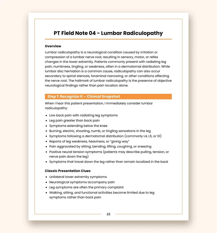
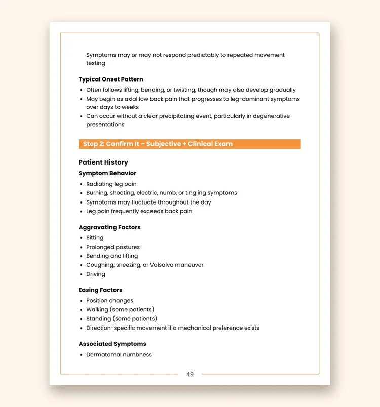
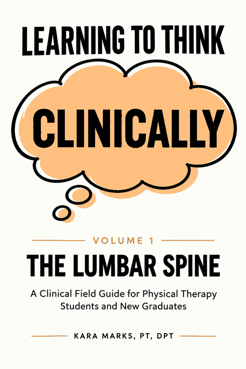
These roadmaps are the short version. The complete shoulder field notes are coming in Learning to Think Clinically, Volume 2: The Shoulder. While you wait, Volume 1: The Lumbar Spine shows you exactly what each field guide looks like inside.
- Every field note, with the complete examination sequence
- The "why" behind each exam and treatment decision
- Differential diagnosis, red flags, and when to refer
- Evaluation templates, clinical cases, and a full clinical reference appendix

Evaluation Templates
Nine fillable PDF templates that walk you through the full exam, from subjective history to assessment and goals.
Get the free templatesA quick note
Clinical Roadmaps are for educational purposes for students and clinicians. They are not a substitute for an individualized evaluation and plan of care from a licensed physical therapist.